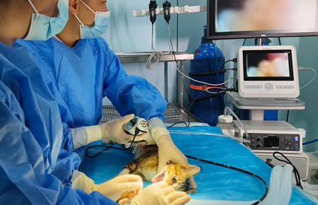
How to handle, care for and prepare a veterinary endoscope?
Veterinary endoscopy has become an invaluable tool in modern diagnostics and surgical interventions for animals. Proper handling, care, and preparation of endoscopic equipment are essential to ensure optimal performance, enhance patient safety, and prolong the lifespan of these sophisticated instruments. This blog outlines best practices for veterinary professionals working with JeetVet endoscopes.

1. Understanding the Endoscope
Before handling an endoscope, it is crucial to familiarize yourself with its components. Typically, a veterinary endoscope consists of the following parts:
Insertion Tube: The flexible or rigid tube that is inserted into the patient.
The insertion tube is connected to the distal aspect of the endoscope body. This is the working end of the scope (i.e. the tube which will enter the patient). It usually has pre-marked 5-10cm measurements on the outside of the tube, so the clinician can easily identify how far in they are. The insertion tube is incredibly delicate and contains the instrument channel, air/water and suction channels, angulation wires.
The distal end of the insertion tube contains the bending section and the distal tip. The bending section is where tip deflection (moving the insertion tube, in order to drive through body channels) occurs. Steel wires move the end of the tube in line with the angulation dials on the scope body. These wires can stretch and break over time and so the angulation should always be operated carefully.
Optical System: Contains lenses that provide high-quality images of internal structures.
Working Channel: Allows for the introduction of tools such as forceps or biopsy instruments.

A gastroscope is generally the largest, longest endoscope used in practice. This has an umbilical tube as well as an insertion tube, contains an air/water channel, larger instrument channel, and has 4-way angulation (the bending section can move left and right, as well as up and down).
Compared with the traditional endoscopes, JeetVet veterinary gastroscopes are innovative design with 360° omnidirectional rotation, allowing for flexible operation and a wider field of view with clear high-definition image display.
The portable veterinary gastroscope is used to examine the esophagus, stomach, duodenum and colon. Some practices have a colonoscope, which has exactly the same functions as a gastroscope but has a slightly longer insertion tube.
2.Handling the Endoscope
As we know, endoscopes are delicate and that can make handling them a little fear-inducing! However, as long as we handle them safely and carefully, we’ll minimise the risk of any damage.
a. Pre-Use Checks:
Inspect the endoscope for any visible damage, such as cracks in the insertion tube or broken optical components.
Ensure that all connections are secure and functioning properly.
b. Clean Hands:
Always handle the endoscope with clean, gloved hands to prevent contamination. If gloves become soiled, change them immediately.
c. Gentle Manipulation:
Avoid excessive force when inserting or maneuvering the endoscope within the patient. Gentle, controlled movements help prevent damage to both the instrument and the animal's tissues.

3. Caring for the Endoscope
a. Cleaning Protocol:
Follow a strict cleaning protocol immediately after use. This typically includes rinsing the endoscope with sterile saline and wiping it down with a soft, lint-free cloth.
Use enzymatic cleaners specifically designed for endoscopic instruments to remove organic debris. Avoid harsh chemicals that may damage sensitive components.
b. Disinfection:
After initial cleaning, disinfect the endoscope using appropriate solutions, following manufacturer guidelines.
c. Drying and Storage:
Thoroughly dry the endoscope before storage to prevent moisture-related damage and microbial growth. Utilize a designated drying cabinet if available.
Store the endoscope in a protective case or holder to prevent accidental damage. Ensure the storage area is clean and free from contaminants.
Always store your endoscopes hanging up, on a rack or wall holder. Most endoscopy trolleys either come with a holder, or one can be added to them. Once the endoscope is set up and ready for use, it should be stored hanging vertically, rather than on a table or work surface. This minimises the risk of drop/fall injuries, as well as things coming in to contact with the scope.
4. Preparing the Endoscope for Use
Pre-Procedure Setup:
Ensure that all components are functioning correctly before the procedure.
Perform a final check on the working channel to confirm it is clear and free from obstructions. Test the optical system to verify image quality.
Documentation:
Maintain a log of endoscope use, including cleaning and maintenance records. This helps ensure compliance with veterinary standards and assists in tracking the instrument's performance over time.

Conclusion
Proper handling, care, and preparation of veterinary endoscopes are vital to ensure successful diagnostic and therapeutic outcomes. By following these best practices, veterinary professionals can enhance the longevity of their instruments while providing safe and effective care for their patients. As technology continues to advance, staying informed about the latest techniques and recommendations will further improve endoscopic procedures in veterinary medicine.
Looking for reliable, high-quality endoscopy equipment? Visit JeetVet.com to discover our range of veterinary endoscopes designed for precision and ease of use.


