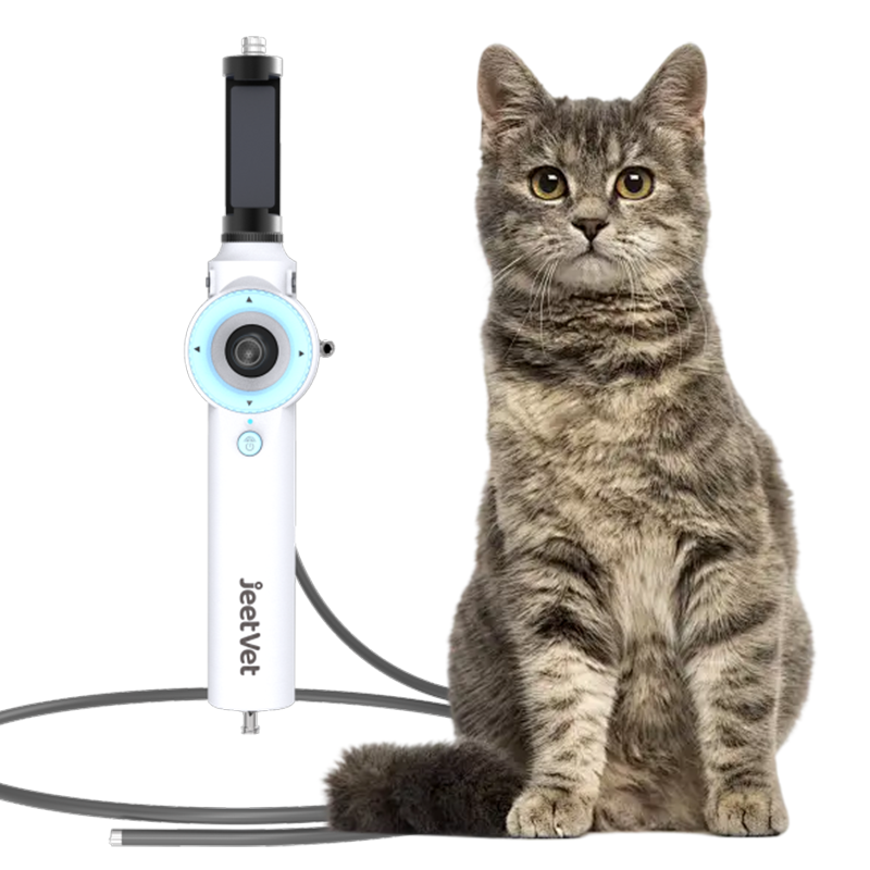
Common GI Disorders in Animals Diagnosed with Veterinary Endoscope
Gastrointestinal (GI) disorders are a common concern in veterinary medicine, affecting animals of all ages and breeds. Portable veterinary endoscope has become an indispensable tool for diagnosing these conditions quickly and non-invasively. In this article, we will explore common GI disorders in animals identified through upper and lower GI endoscopy, highlighting procedures like esophagoscopy, gastroscopy, duodenoscopy, and colonoscopy.

Upper Gastrointestinal (GI) Endoscopy: Diagnosing Esophageal, Stomach, and Duodenal Issues
Upper GI endoscopy involves inserting a flexible veterinary endoscope through the mouth to examine the esophagus, stomach, and duodenum. Key applications include:
1. Esophagoscopy
Esophagoscopy is used to diagnose and treat esophageal diseases such as:
Esophageal strictures: Narrowing of the esophagus due to scar tissue or inflammation.
Foreign bodies: Objects like bones or toys lodged in the esophagus.
Ulcers: Erosions or sores in the esophageal lining caused by acid reflux or trauma.
Why It Matters: Enables direct visualization and retrieval of obstructions, reducing the need for invasive surgery.
2. Gastroscopy
Gastroscopy allows veterinarians to examine the stomach and retrieve foreign objects non-surgically. Common conditions diagnosed and treated include:
Gastric ulcers: Open sores in the stomach lining.
Stomach tumors: Abnormal growths that may require biopsy or removal.
Foreign body retrieval: Removing objects like toys or bones from the stomach.
Key Advantage: Allows non-surgical removal of foreign objects from the stomach using usb veterinary gastroscope with endoscopic graspers.
3. Duodenoscopy
Duodenoscopy focuses on the duodenum (the first part of the small intestine). It helps diagnose conditions such as:
Inflammatory bowel disease (IBD): Chronic inflammation of the intestinal lining.
Tumors: Abnormal growths that may obstruct the duodenum.
Blockages: Physical obstructions caused by foreign bodies or strictures.
Clinical Value: Facilitates biopsies to differentiate between IBD, infections, or cancer.

Lower Gastrointestinal (GI) Endoscopy: Evaluating the Colon and Ileum
Lower GI endoscopy (colonoscopy/ileoscopy) involves inserting the animal endoscope through the rectum to assess the colon and ileum. It helps diagnose and treat conditions such as:
1. Colonoscopy
Colitis (inflammation due to parasites, infections, or allergies).
Colonic tumors (e.g., benign polyps or malignant growths).
Chronic diarrhea of unknown origin.
Procedure Insight: Biopsies collected during colonoscopy help identify causes of persistent diarrhea or bleeding.
2. Ileoscopy
Ileal inflammation (often linked to IBD).
Obstructions or strictures in the ileum.
Infections (e.g., bacterial overgrowth or parasitic infestations).
Benefits of Gastroscopy for GI Diagnosis in Animals
1)Minimally Invasive: Veterinary endoscopic procedures cause minimal discomfort and tissue damage to the animal, shortening the animal's recovery time and allowing the animal to return to normal activities more quickly.
2)High Precision: Endoscopes can visualize the internal structures of the animal in real time, allowing veterinarians to identify abnormalities promptly and accurately.
3)Biopsy and Treatment: Tissue samples can be collected, and treatments such as removal of foreign objects or application of medication can be performed.

Conclusion
Veterinary Endoscope is a versatile and minimally invasive tool for diagnosing and treating gastrointestinal disorders in animals. By understanding the applications of upper and lower GI endoscopy, veterinarians can provide early intervention and improve patient outcomes.
Ready to enhance your veterinary practice with advanced endoscopy solutions? Visit our website JeetVet.com today to explore our range of high-quality veterinary endoscopy equipment designed for precision and ease of use.


