
A Comprehensive Guide to Using Veterinary Endoscopes for Feline Patients
Veterinary endoscopy has revolutionized the way veterinarians diagnose and treat various medical conditions in feline patients. This minimally invasive technique allows for direct visualization of internal structures, collection of tissue samples, and even therapeutic interventions with minimal discomfort to the patient. Given the delicate nature of feline anatomy and their propensity for stress-related complications, the use of endoscopy in cats requires a high level of precision, expertise, and understanding of the equipment. This blog aims to provide a detailed, professional guide on how to effectively use portable animal endoscopes for feline patients, covering indications, preparation, techniques, and post-procedural care.

Indications for Endoscopy in Feline Patients
Endoscopy is indicated in feline medicine for both diagnostic and therapeutic purposes. Common diagnostic indications include:
1. Gastrointestinal Disorders: Chronic vomiting, diarrhea, weight loss, and anorexia may warrant endoscopic evaluation of the esophagus, stomach, and duodenum. Endoscopy allows for the identification of conditions such as inflammatory bowel disease, neoplasia, and foreign bodies.
2. Respiratory Conditions: Nasal discharge, sneezing, and stridor may necessitate rhinoscopy or bronchoscopy to assess the nasal passages, sinuses, and airways for inflammation, polyps, or tumors.
3. Urinary Tract Disorders: Cystoscopy is employed to evaluate hematuria, dysuria, and recurrent urinary tract infections. It is particularly useful for diagnosing conditions such as bladder stones, tumors, and urethral obstructions.
4. Ear and Ocular Conditions: Otoscopy is used to examine the ear canal for infections, polyps, or foreign bodies, while ophthalmoscopy aids in the evaluation of ocular structures.
Therapeutic applications of endoscopy in cats include the removal of foreign bodies, polypectomy, balloon dilation of strictures, and placement of feeding tubes.
Preparation for Endoscopic Procedures
Proper preparation is critical to the success of endoscopic procedures in feline patients. The following steps should be taken:
1. Patient Evaluation: A thorough physical examination, along with diagnostic tests such as blood work, radiographs, and ultrasound, should be performed to assess the patient’s overall health and identify any contraindications to anesthesia.
2. Fasting: To ensure optimal visualization and reduce the risk of aspiration, cats should be fasted for 12 hours prior to gastrointestinal endoscopy. Water may be withheld for 2-4 hours before the procedure.
3. Anesthesia: Endoscopy in cats requires general anesthesia to ensure patient immobility and comfort. A pre-anesthetic assessment should be conducted, and appropriate anesthetic protocols tailored to the individual patient’s needs should be implemented. Endotracheal intubation is recommended to protect the airway, especially during upper gastrointestinal or respiratory endoscopy.
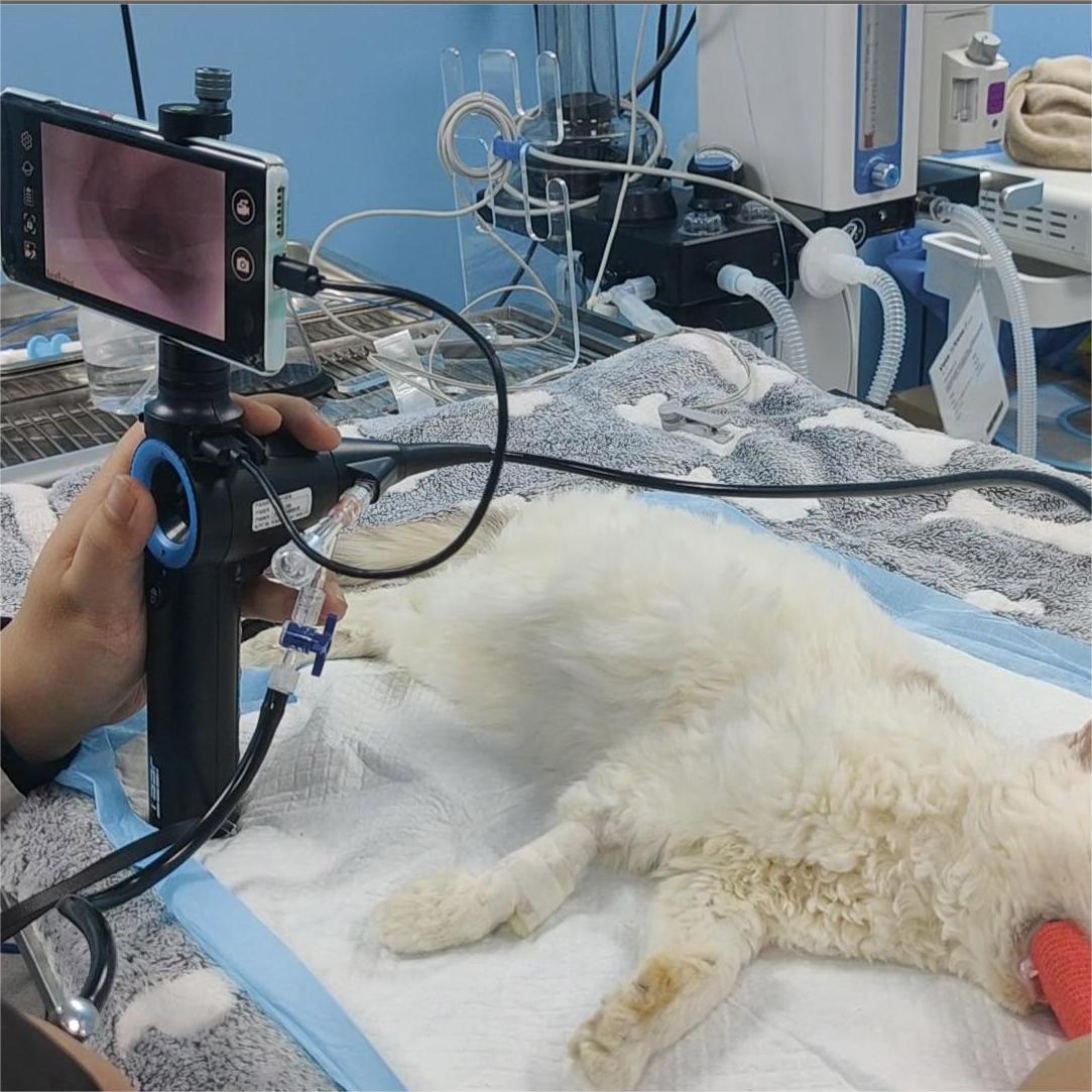
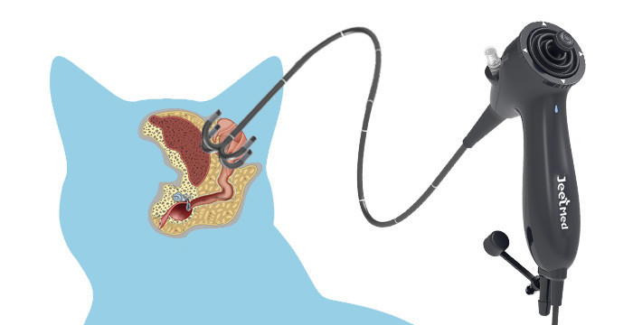
4. Equipment Preparation: The animal endoscope should be thoroughly cleaned and sterilized according to manufacturer guidelines. Ensure that all necessary accessories, such as biopsy forceps, grasping tools, and suction devices, are readily available. A video endoscopy system is preferred for its superior image quality and ability to record findings for further analysis.
Post-Procedural Care
Post-procedural care is essential to ensure the patient’s recovery and minimize complications. Key considerations include:
Monitoring: Cats should be closely monitored during recovery from anesthesia. Vital signs, including heart rate, respiratory rate, and temperature, should be assessed regularly. Oxygen supplementation may be necessary in some cases.
Pain Management: Although endoscopy is minimally invasive, some patients may experience discomfort. Appropriate analgesics should be administered as needed.
Dietary Management: After gastrointestinal endoscopy, cats should be offered a small amount of water once fully awake. A bland diet may be introduced gradually, with a return to normal diet over 24-48 hours.
Complication Management: Complications such as bleeding, perforation, or infection are rare but possible. Any signs of distress, such as vomiting, lethargy, or difficulty breathing, should be promptly addressed.


